In the cold depths of the ocean, nature is slow to give up its secrets. In April, scientists discovered the first adult white orca in the wild. Following on the heels—or tail—of this find, a group of other researchers have identified a new sensory organ in a group of baleen whales.
The newly discovered organ helps explain how the whales lunge feed, a method they rely upon to maintain their enormous size. The sensory organ also provides insight into the whales’ evolutionary history.
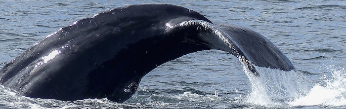
Scientists from the Smithsonian Institution and the University of British Columbia examined tissues collected during commercial whale catches. They focused their efforts on rorqual whales, which include fin, humpback, and blue whales.
In spite of hundreds of thousands of whales being collected during catches over the past decades, scientists are “still only beginning to understand the anatomy of the largest ocean predators of all time,” said Nicholas Pyenson, paleobiologist at the Smithsonian’s National Museum of Natural History and lead author of the research, in a press release.
Lunge Feeding
 Among the largest vertebrates on the planet, rorqual whales—like the blue whale—can grow to more than 100 feet long and weigh more than 150 tons. They accomplish this entirely by eating some of the smallest organisms in the ocean.
Lunge feeding provides them with the vast amount of food needed to maintain their enormous size. The whales race forward and take in more water than their own body weight. When they clamp their jaws shut, they filter out the millions of krill and small fish that make up their diet.
Until the discovery of the sensory organ, scientists were uncertain exactly how the whale coordinated the many parts involved in successful lunge feeding. It turns out that the organ is ideally situated for its function.
Structure and Function of the Sensory Organ
Located in the chin of rorqual whales, the organ is pinched between the tips of the two bones that make up the lower jaw. It consists of several kinds of tissue, including blood vessels, nervous tissue with sensory receptors, and a gel-like cavity.
The sensory organ responds to movements in the jaw that occur when the whale opens and closes its mouth. It also picks up on the expansion of the throat pleats that happens when the whale takes in water.
During lunge feeding, the tips of the jaw pinch the organ. Nerves that connect it to the brain sense the changes in the shape of the organ. This tells the whale to close its jaw before its prey can escape.
This new evidence shows that “rorquals actively control their lunge, rather than letting their mouths passively inflate like a parachute,” said Pyenson.
Evolution of the Sensory Organ
In addition to illuminating the feeding mechanisms of rorqual whales, the sensory organ also provides scientists with insight into the evolutionary history of the whales.
The fossil record shows that the bottom jaw of baleen whales—like the rorquals—has been separated or unfused at its tip for 23 to 28 million years (since the late Oligocene epoch).
The sensory organ sits in an ancestral tooth socket that is still present in rorqual whales today. The organ, though, is an evolutionary novelty for rorqual whales. It doesn’t appear in other modern species of baleen whales, like the right and gray whales.
To date, scientists have only identified this type of structure in the rorqual whales, giving them a special place in the ocean alongside the infamous white orca.
One more mystery of the deep revealed, but many more remain.
__________
Reference: Pyenson, N., Goldbogen, J., Vogl, A., Szathmary, G., Drake, R., & Shadwick, R. (2012). Discovery of a sensory organ that coordinates lunge feeding in rorqual whales Nature, 485 (7399), 498-501 DOI: 10.1038/nature11135
Photo: Scientists from the University of British Columbia and the Smithsonian Instiution point to a ridge of tissue sampled from the throat pouch a fin whale (background) in Iceland. Left to right: Jeremy A. Goldbogen (Cascadia Research Collective), A. Wayne Vogl (UBC) and Robert E. Shadwick (UBC). Photo credit: Nicholas D. Pyenson / Smithsonian Institution.